


Computers can do this kind of calculation very fast, but using a computer in an operating room in the early 1970's was almost unheard of!!!

Early uses of computer generated brain maps for use in Stereotaxic Surgery.
1. Thompson C J and Bertrand G: "A computer program to aid the neurosurgeon to locate probes used during stereotaxic surgery on deep cerebral structures". Comp Prog in Biology 2: 265-276 (1972)
2. Bertrand G, Olivier A and Thompson C J: "Computer display of stereotaxic brain maps and probe tracts". Acta Neurochir (Suppl) 21: 235-243 (1974)
3. Thompson C J, Hardy T and Bertand G: "A system for anatomical and functional mapping of the human thalamus." Comput Biomed Research 10: 9-24 (1977)
|
|
|
|
| In the 1970's Stereotaxic surgery was widely used in the treatment of Parkinson's disease. A frame like the one on a skull above is attached to the patient's head and probes can be directed to deep structures in the brain, like the thalamus which serve as relays and are vital in normal movement and coordination. In those days, before the invention of CT scanning and MRI it was impossible to "see" the brain with X-rays as they are attenuated by the skull. X-ray opaque dye was injected into the brain's ventricles in order to locate them with respect to the frame, and maps were then used to locate structures as surgical targets. | Stereotaxic surgery relies on making lesions (cuts in the brain) with a small tool at the end of a long probe, and it is essential to know where the probe is before making the lesion as it can't be reversed. From the engravings on base of the frame and movable arcs, the angles and position can be measured relative to the skull. But not all brains are the same size, so as well as the complex 3D geometry, some scaling is required to match the patient's brain to a reference brain atlas |
This set of equations represents the coordinate transformation to go from
the reference position of the probe tip P to the current
position P'. This is not the sort of thing one would expect a
surgeon to do in his head during such delicate and non-reversible surgery! Computers can do this kind of calculation very fast, but using a computer in an operating room in the early 1970's was almost unheard of!!! |
|
|
|
|
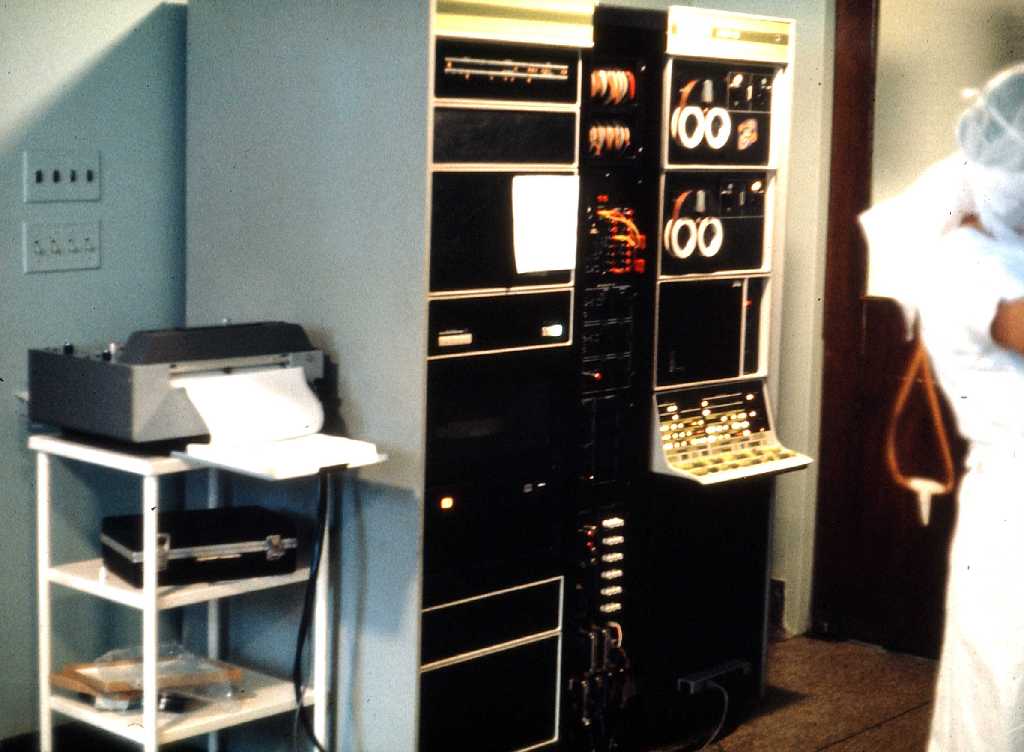
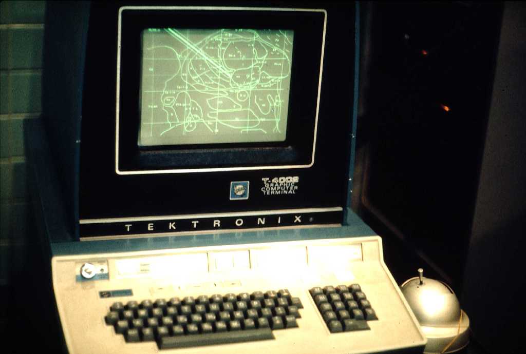
| This picture shows the PDP-12 computer outside the operating room during a Stereotaxic procedure in 1972. A plotter on the left made paper copies of the maps displayed of the terminal screen. The cabinet on the left contained a removable disk drive which stored digitized maps from a reference brain. The disk capacity was 1.2 Mbytes! The processor, with 16 Kbytes of memory was in the right cabinet. The CPU speed was 500 KHz! The nurse on the right and all other personnel left the operating room during the times verification X-rays were made to show the probe's location. | The maps, scaled to the present patient, were displayed on this Tektronix graphics terminal, placed next to the surgeon. It had a storage screen with a resolution of 1024x768 pixels, but no grey-scale. It took about 3-5 seconds to display an new map and probe on the screen while doing the calculations shown above on every point displayed. The "joystick" at the right was used as a pointer on the screen much like a mouse is used today. This was the first terminal of this type used in Canada, and two engineers came from Oregon to install it! The terminal cost over $5,000. | This image shows clusters of letters overlaid on a map of the central brain structures. Each letter was a code for a cell from which a micro-electrode recording was made during any of the Stereotaxic procedures. Data from all patients was pooled after scaling the brains to a common size. By selecting only those units which responded to, (for example): "touching the right side of the upper lip", one could observe clusters of the corresponding letter, giving some indication of the individual variability of the location of these sensory cell. |