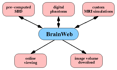
next_inactive up previous


BrainWeb:
Online Interface to a 3D MRI Simulated Brain Database


CHRIS A. COCOSCO, VASKEN KOLLOKIAN, REMI K.-S. KWAN, G. BRUCE PIKE, ALAN C. EVANS

McConnell Brain Imaging Centre, Montréal Neurological Institute,

McGill University, Montréal, Canada


Introduction:

The increased importance of automated computer techniques for anatomical brain mapping from MR images and quantitative brain image analysis methods leads to an increased need for validation and evaluation of the effect of image acquisition parameters on performance of these procedures. Validation of analysis techniques of in-vivo acquired images is complicated due to the lack of reference data (``ground truth''). Also, optimal selection of the MR imaging parameters is difficult due to the large parameter space. BrainWeb makes available to the neuroimaging community, online on WWW, a set of realistic simulated brain MR image volumes (Simulated Brain Database, SBD) that allows the above issues to be examined in a controlled, systematic way.

Methods:

The 3D simulated MR images are generated by varying specific imaging parameters and artifacts in an MRI simulator, which:

$\bullet$
starts from a fuzzy digital phantom containing the spatial probabilistic distribution of different tissue types [1]; as an example of the generality of this approach, other brain phantoms were obtained by adding MS lesions (extracted from real MRI-s) to the normal phantom (Fig. 1).
$\bullet$
computes signal intensities based on Bloch equations or signal equations, accounting for partial volume according to the scan parameters [2] (Fig. 2)
Image noise was specified as a percent standard deviation relative to the mean signal intensity for a reference brain tissue. Multiplicative receive intensity non-uniformity (INU) was simulated using fields recovered from real scans, and scaled for different percent sensitivity ranges.


  \psfig{figure=figs/phantom_1.0mm_normal_wht_t90.eps,width=0.225\linewidth}   \psfig{figure=figs/phantom_1.0mm_normal_gry_t90.eps,width=0.225\linewidth}   \psfig{figure=figs/phantom_1.0mm_normal_csf_t90.eps,width=0.225\linewidth}   \psfig{figure=figs/phantom_1.0mm_msles3_wml_t90.eps,width=0.225\linewidth}  


$\textstyle \parbox{0.9\linewidth}{ \figurenum example of fuzzy tissue classes
from the phantom: (L to R) white matter, gray matter, CSF, MS lesions}$


\psfig{figure=figs/real_vs_sim.eps,width=0.9\linewidth}

$\textstyle \parbox{0.9\linewidth}{
\figurenum real (top) and simulated (bottom) MRI-s (both INU corrected)}$


Features:


\psfig{figure=figs/brainweb.eps,width=0.85\linewidth}

$\textstyle \parbox{0.9\linewidth}{
\figurenum BrainWeb features overview
}$


A pre-computed SBD was generated by varying specific imaging sequence and artifact parameters. The set of these of parameters was chosen according to the values typically encountered in modern MRI 
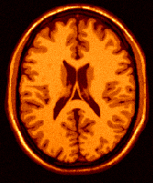
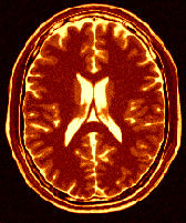
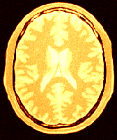
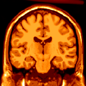
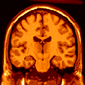
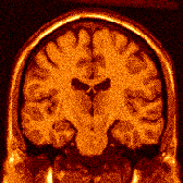
systems [3]. For two anatomical models (normal, and with moderate MS lesions), simulated volumes for three imaging sequences are available online (T$_1$, T$_2$, PD), each with several 
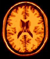
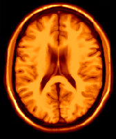
values of slice thickness, noise and intensity INU levels
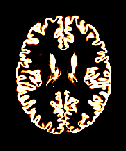
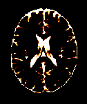
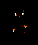
 (Fig. 4).


\psfig{figure=figs/ns_sbd_selection.ps,width=0.75\linewidth}

$\textstyle \parbox{0.9\linewidth}{
\figurenum WWW page: SBD selection for the pre-computed volumes
}$

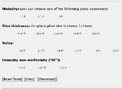
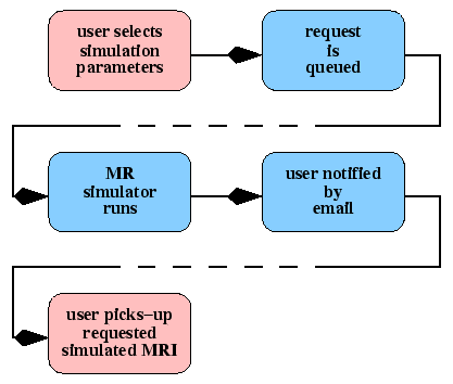
In addition, BrainWeb allows a remote user to run their own custom MRI simulation (on our server) with any of several pulse sequences and source digital phantoms, and arbitrary values of the acquisition artifacts (Figs. 56). After the simulation is finished, the user can preview the result online or download it, everything being done through the WWW interface.


\psfig{figure=figs/mrisim_ctrl.eps,width=0.75\linewidth}
$\textstyle \parbox{0.9\linewidth}{
\figurenum generating custom MRI simulations
}$


acquisition parameter selectable values
phantom normal;
  MS lesion mild, moderate, severe
  slice thickness 1 - 10 mm
  scan technique SE, IR, SFLASH, CEFAST, FISP,
    FLASH, DSE_EARLY, DSE_LATE
MR pulse sequence repetition time (any) ms
  inversion time (any) ms
  flip angle 1 - 150 deg
  echo times (any) ms
  partial volume (slice thickness) 1 - 10 mm
imaging artifacts noise 0 - 10 %
  INU 0 - 60 %


$\textstyle \parbox{0.9\linewidth}{
\figurenum parameters that can be controlled for custom MRI simulations
}$

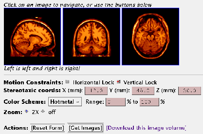
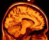
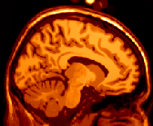
All 3D image volumes are in stereotaxic space, and can be interactively explored with practically any WWW browser in three simultaneous orthogonal views at any cursor position in the volume (Fig. 7). Each simulated brain image, as well as the source digital phantoms, can be downloaded in a variety of file and data compression formats.


\psfig{figure=figs/ns_bw_view.ps,width=0.75\linewidth}

$\textstyle \parbox{0.9\linewidth}{
\figurenum WWW page: BrainWeb online viewer
}$


Applications:

SBD can be used to:

$\bullet$
study the performance of anatomical brain mapping techniques (as a function of different MR acquisition parameters), such as: non-linear co-registration [4], cortical surface extraction [5], or tissue classification algorithms [3].
$\bullet$
validate quantitative analyses of neuropathology (e.g. MS lesion quantification [6]), or other medical pattern recognition and image processing techniques.
The main advantages of using these simulations are:
$\bullet$
the answer is known a priori in the experiment.
$\bullet$
parameters can be independently controlled (Figs. 8, 9, 10, 11).
Since the source for all simulations is the same digital phantom, one has a systematic means of establishing the sensitivity of any particular procedure with respect to any imaging parameter or artifact.


  \psfig{figure=figs/t1_icbm_normal_1mm_pn3_rf20_t90.eps,width=0.3\linewidth}   \psfig{figure=figs/t2_icbm_normal_1mm_pn3_rf20_t90.eps,width=0.3\linewidth}   \psfig{figure=figs/pd_icbm_normal_1mm_pn3_rf20_t90.eps,width=0.3\linewidth}  


$\textstyle \parbox{0.9\linewidth}{
\figurenum T$_1$, T$_2$, PD weighted (typical noise and INU levels)}$


  \psfig{figure=figs/t1_icbm_normal_1mm_pn0_rf0_c108.eps,width=0.3\linewidth}   \psfig{figure=figs/t1_icbm_normal_1mm_pn3_rf0_c108.eps,width=0.3\linewidth}   \psfig{figure=figs/t1_icbm_normal_1mm_pn9_rf0_c108.eps,width=0.3\linewidth}  


$\textstyle \parbox{0.9\linewidth}{
\figurenum Noise (but no INU): zero, typical (3\%), extreme (9\%)}$


  \psfig{figure=figs/t1_icbm_normal_1mm_pn0_rf0_s80.eps,width=0.3\linewidth}   \psfig{figure=figs/t1_icbm_normal_1mm_pn0_rf20_s80.eps,width=0.3\linewidth}   \psfig{figure=figs/t1_icbm_normal_1mm_pn0_rf40_s80.eps,width=0.3\linewidth}  


$\textstyle \parbox{0.9\linewidth}{
\figurenum INU (but no noise): zero, typical (20\%), extreme (40\%)}$


  \psfig{figure=figs/t1_icbm_normal_1mm_pn0_rf0_t90.eps,width=0.3\linewidth}   \psfig{figure=figs/t1_icbm_normal_5mm_pn0_rf0_t18.eps,width=0.3\linewidth}   \psfig{figure=figs/t1_icbm_normal_9mm_pn0_rf0_t10.eps,width=0.3\linewidth}  


$\textstyle \parbox{0.9\linewidth}{
\figurenum slice thickness and partial volume effect: (L to R) 1mm,
5mm, 9mm slices (no noise, no INU)}$


Conclusion:

BrainWeb provides to the international research community a convenient access to this resource through the Internet. The full SBD, the online interface for controlling the MR simulator, as well as the anatomical models (phantoms) used as input to it, are available on WWW at ``http://www.bic.mni.mcgill.ca/brainweb/''. We are currently working on extending BrainWeb to include fMRI and PET simulated data.

Acknowledgments:

This work supported by the U.S. Human Brain Map Project and the International Consortium for Brain Mapping (ICBM). Contributions to this project were made by Peter Neelin, Alex Zijdenbos, John Sled, and Louis Collins from the McConnell Brain Imaging Centre.

Bibliography

1
Collins, D.L., Holmes, C.J., Evans, A.C.
Human Brain Mapping. 1997

2
Kwan, R.K.-S., Evans, A.C., Pike, G.B.
In Lecture Notes in Computer Science, vol. 1131, pages 135-140, 1996.

3
Kollokian, V.
Master's thesis, Concordia University, Montreal, QC, Canada, Nov. 1996.

4
Collins, D.L., Holmes, C.J., Peters, T.M., Evans, A.C.
Human Brain Mapping. 3(3):190-208, 1996.

5
MacDonald, D., Avis, D., Evans, A.C.
In VBC, Proceedings of the SPIE, vol. 2359, pages 160-169, 1994.

6
Zijdenbos, A.P., Evans, A.C., Riahi, F., Sled, J.G., Chui, H.-C., Kollokian, V.
In Lecture Notes in Computer Science, vol. 1131, pages 439-448, 1996.


About this document ...

This document was generated using the LaTeX2HTML translator Version 2K.1beta (1.49)

Copyright © 1993, 1994, 1995, 1996, Nikos Drakos, Computer Based Learning Unit, University of Leeds.
Copyright © 1997, 1998, 1999, Ross Moore, Mathematics Department, Macquarie University, Sydney.

The command line arguments were:
latex2html -split 0 -mkdir -dir /home/bic/crisco/www/HBM97_poster/ -title 'BrainWeb HBM 1997 Poster' for_html.tex

The translation was initiated by Chris COCOSCO on 2002-07-29


next_inactive up previous
Chris COCOSCO 2002-07-29