The spatial resolution of an imaging modality or instrument denotes its
ability to reproduce fine detail. Ideally, a tomographic image would
exactly reproduce the object or, in the case of nuclear medicine, the
activity distribution within the patient. If the properties of scaling,
superposition, and space invariance are assumed to be preserved to allow
the assumption of a linear system [Web88][BS81a][Mak83], then the
physical response or capability of an imaging system to reproduce the
limit of fine detail, a point source or function, may be
characterized by its point spread response function (PSRF). The stimulus
object is said to have been convoluted or distorted by the system to produce a
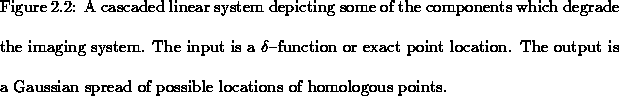
blurry output image. The ``black box'' which represents the entirety of
the imaging system consists of other cascaded linear systems
representing the presence of scatter, attenuation, imperfect electronics,
the apodizing functions used in the tomographic reconstruction and other
factors which all contribute in an imaging system's capability to
reproduce an object exactly [RK82b][RK82a][BS81b][BS81a] (see
figure 2.2).
The degree of blurring introduced as described by the PSRF is most
generally represented by a Gaussian plus a constant pedestal of activity
where the information density follows a Poissonian distribution according
to nuclear decay statistics (see figure 2.3). Better spatial
resolution, or a narrower Gaussian distribution, may be obtained in
practice by compensating for scatter and the depth dependent detector
response in SPECT systems with the use of iterative filtered back
projections and maximum a posteriori probability procedures
[MCC+93][KZR92][Lia93]. The use of elliptical scans which more closely
approximates the transaxial outline of the head also improves the spatial
resolution by minimizing the source to detector distance. Attenuation
correction studies [KPD+92] using simple first order techniques like
the Sorenson hyperbolic sine method [Sor74][Kem89] have shown that
the effect of attenuation on the spatial resolution is insignificant; it
is nevertheless an important correction for quantitative studies. Methods
which correct for the response of the different components in the cascaded
linear systems are non-trivial because of the computational intensity
required for their implementation (of the order of hours for typical
128128
128 image arrays) [Lia93] and, as such, are
often not clinically utilised.
The extension of the PSRF to a volume data set is naturally a 3-D
Gaussian envelope. The width of the Gaussian on top of the background of
noise in each dimension may be described by standard deviation.
A good imaging system is said to have a narrow PSRF so that the spatial
position of the original point source may be more accurately determined
from information from the image alone. The dimensional components of the
3-D PSRF in image space are generally not equal. In figure 2.4
depicting point sources on and
off the center of rotation in a tomographic plane perpendicular to the
axis of rotation, the shape of the circular cross section of a point
source tends to be stretched out in the radial direction to a more
elliptical shape. Note that the x and y dimensions depicted in the figure
are usually oriented from left to right and from the bottom to the top,
respectively. In terms of the anatomical orientation of the brain, this
corresponds to the neurological convention [KW92] of viewing
transaxial cuts where top is the anterior, bottom is the
posterior, left is the patient's left, etc. (please see the
appendix). The radial component of the PSRF off axis tends to become
greater than the tangential component to ultimately give this distorted
oblong shape [SP87][OFBM88][UKMM81][Gre83][KPD+92]. This is due to increased
rejection of large angle scatter by the collimator septa as the source
moves farther away from the center of rotation and closer to the detector
face (see figure 2.4). This is the same cause of resolution
improvement which is achieved by using elliptical scans, as mentioned
above, because the activity distribution within a patient is brought
closer to the detector face. For an elliptical scan then, points radially
further away from the axis of rotation along the major axis and in the
plane perpendicular to the axis of rotation (usually the transaxial slice)
will generally have diminished resolution. For scans which revolve about
the inferior-superior axis, the orientation of diminished resolution
therefore correspond to the anterior-posterior direction because the
brain's dimension is largest in this direction.
