In addition to anatomical information derived from MRI, MRA, CT and DSA, it is vitally important for the surgeon to have an appreciation of the functional activity of critical regions. The need could be to determine areas of low or zero metabolic activity that could be safely removed, or conversely to determine the precise location of sensory or motor regions in the vicinity of a surgical site in order that they might be spared. The objective here was therefore to integrate the images derived from PET and MRI to give the surgeon simultaneous access to functional and anatomical information.
It has become standard practice at the MNI to acquire an MRI scan
of each patient undergoing a PET study. In addition, methodology is
now well established to enable the 3-D images from each of the
modalities to be combined[15][16]. We use a
landmark-matching algorithm that minimizes the mean square distance
between homologous points identified in each of two
volumes[17]. Using a workstation display that allows
interactive manipulation and display of transverse, sagittal and
coronal views from each data set, the operator identifies a set of
(typically 20) homologous points in the two data sets. This
procedure (known as the Procrustes algorithm) is now in routine
use to correlate each PET data set with its corresponding
MRI volume with a registration accuracy of
2-3mm[15].
Our implementation of PET/MRI imaging on the surgical workstation, consists of a tri-planar display showing orthogonal slices of combined images, along with a 3-D display of object surfaces derived from the MRI and PET images. The PET activity in each slice is indicated by the colour scale superimposed on the underlying grey-scale MRI image. The relative contribution of each data set to the final image is under the interactive control of the operator. The position of the surgical probe in relation to each data set is indicated by a cross-hair cursor on each of the tri-plane images, as well as by its realistic representation in the 3-D image. Fig. 4 shows typical merged MRI and PET data, in this case for a patient undergoing surgery to remove cerebral tissue during the treatment of intractable epilepsy. The PET and MRI are clearly visible in the orthogonal slices of the triplanar display. The bottom right section of the screen shows surface rendering of segmented 3-D objects the probe (purple), skin (white), cortex (red), and a PET activation level (yellow) that was chosen to yield information relevant for this particular procedure. In order to enhance the realism of the 3-D images, they are displayed in stereo on demand.
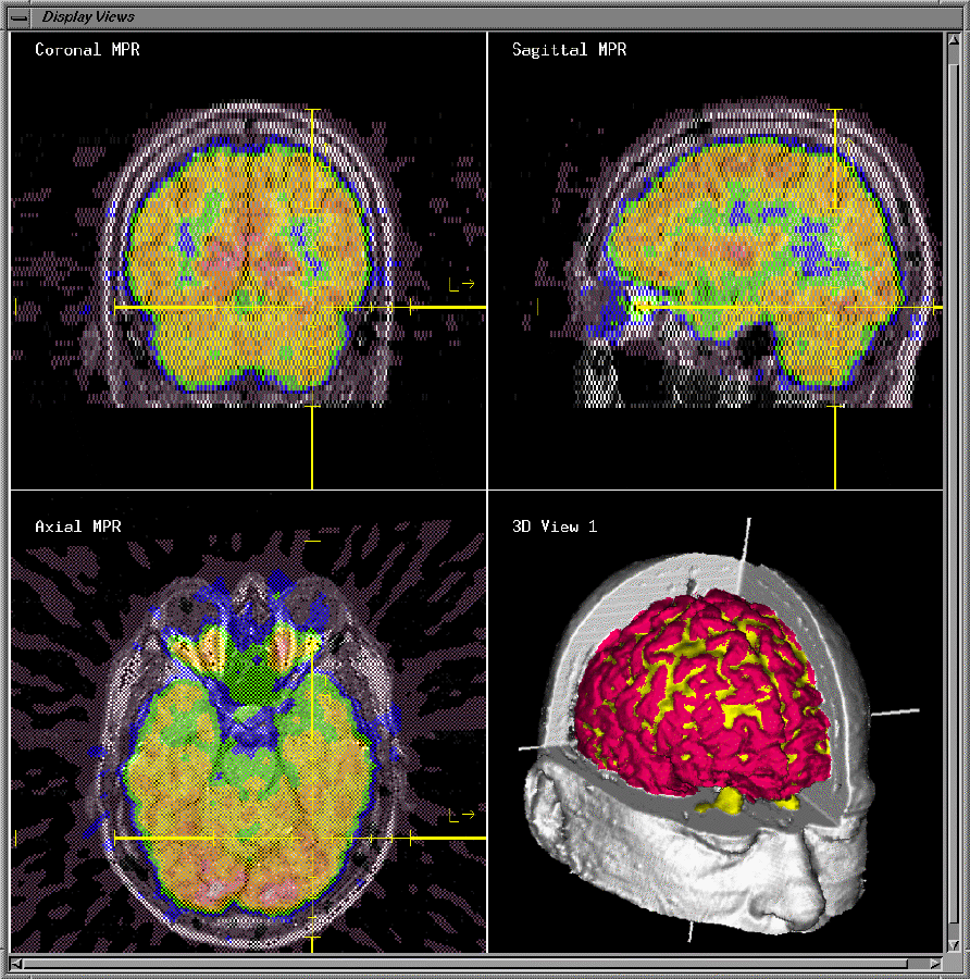 Figure 4: Click on image.