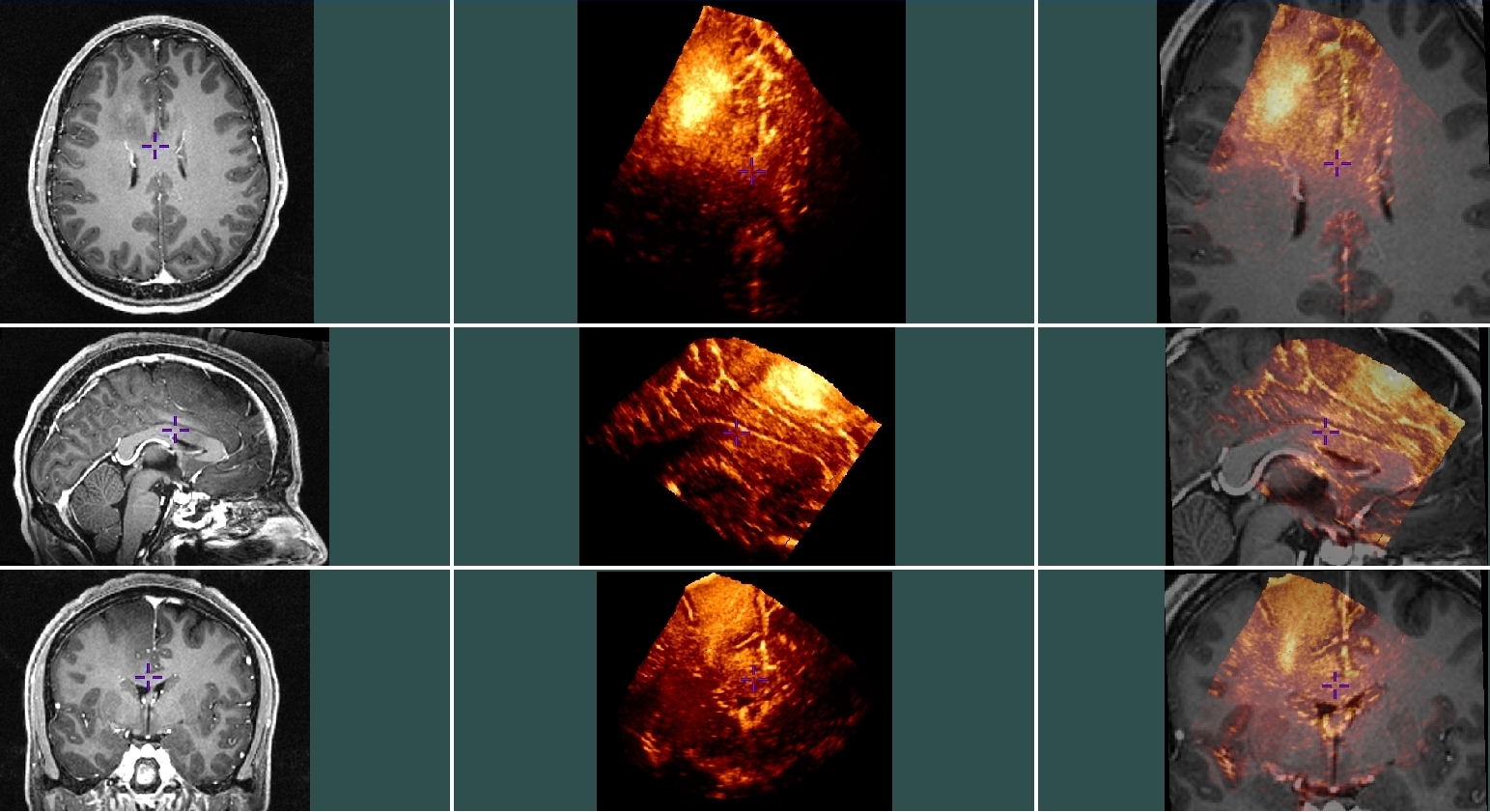
Group 2 image example: case #8
back to data page
Left: Pre-operative T1-weighted MRI with gadolinium (grey tones)
Center: Pre-resection ultrasound (hot tones)
Right: The two images combined (note that manual tags were used to better align the two images for an easier visualization).