Abstract
Objective: To determine the relationship between hippocampal, amygdalar, and entorhinal cortex atrophy and duration of epilepsy, presence of secondary generalized seizures, and prolonged childhood febrile convulsions in patients with pharmacologically intractable temporal lobe epilepsy (TLE).
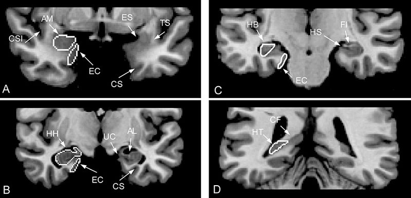
Methods: Volumetric MRI of the hippocampus, amygdala, and entorhinal cortex were performed in 86 consecutive patients with TLE and 44 age- and sex-matched healthy control subjects. Linear regression analysis was used to explore the relation between the volumetric measurements and the clinical parameters.
Results: In simple regressions, duration of epilepsy but not age at seizure onset was related to hippocampal (r2 = 0.19, p < 0.001), entorhinal cortex (r2 = 0.11, p = 0.002), and amygdalar (r2 = 0.15, p < 0.001) atrophy ipsilateral to the seizure focus. Prediction of the regression function to time of onset of recurrent seizures (time = 0) resulted in a y intercept of < 0 for the hippocampus and the entorhinal cortex but was not different from 0 for the amygdala. Patients with a positive history of febrile convulsions had smaller hippocampal volumes ipsilateral to the seizure (p < 0.001). No relationship was found between febrile convulsions and entorhinal cortex and amygdalar volumes or between secondary generalization of seizures and any mesial temporal volume.
Conclusion: Progressive volume loss in the mesial temporal lobe in relation to duration of epilepsy is not limited to the hippocampus but affects the entorhinal cortex and the amygdala.