Abstract
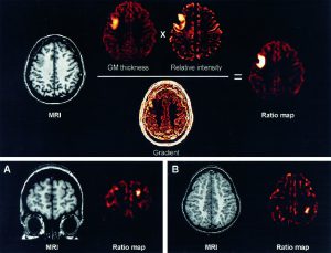
In many patients, focal cortical dysplasia (FCD) is characterized by minor structural changes that may go unrecognized by standard radiological analysis. To increase the sensitivity of magnetic resonance imaging (MRI) for the detection of subtle lesions of FCD, we developed voxel-based image postprocessing methods, including first-order texture analysis and morphological processing modeled on known MRI features of FCD. We selected 16 patients with histologically proven FCD. Image processing features were calculated over a neighborhood for each voxel in the three-dimensional T1-weighted MRI. Three feature maps were generated: (1) gray matter thickness map to model cortical thickening, (2) gradient map to model blurring of the gray matter–white matter junction, and (3) relative intensity map to model the hyperintense signal within the lesion. Two observers detected lesions on conventional MRI in 8/16 and on ratio maps in 14/16 patients. Sensitivity was 87.5% (14/16) for the ratio maps compared to 50% (8/16) for MRI (p < 0.003). Specificity was 95% (19/20) for ratio maps and 100% (20/20) for MRIs. Cohen's kappa was 0.53 for MRIs, indicating moderate agreement, and 0.83 for ratio maps, indicating strong agreement beyond chance between the 2 observers. The image-processing methods developed in this study improve visual detection of FCD, even in cases where no lesion is obvious on MRI. These techniques could increase the number of patients with partial epilepsy who could benefit from surgery.
