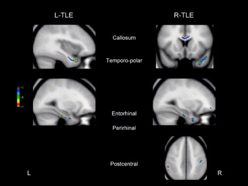
Abstract
Volumetric MRI studies based on manual labeling of selected anatomical structures have provided in vivo evidence that brain abnormalities associated with temporal lobe epilepsy (TLE) extend beyond the hippocampus. Voxel-based morphometry (VBM) is a fully automated image analysis technique allowing identification of regional differences in gray matter (GM) and white matter (WM) between groups of subjects without a prior region of interest. The purpose of this study was to determine whole-brain GM and WM changes in TLE and to investigate the relationship between these abnormalities and clinical parameters. We studied 85 patients with pharmacologically intractable TLE and unilateral hippocampal atrophy and 47 age- and sex-matched healthy control subjects. The seizure focus was right sided in 40 patients and left sided in 45. Student's t test statistical maps of differences between patients' and controls' GM and WM concentrations were obtained using a general linear model. A further regression against duration of epilepsy, age of onset, presence of febrile convulsions, and secondary generalized seizures was performed with the TLE population. Voxel-based morphometry revealed that GM pathology in TLE extends beyond the hippocampus involving other limbic areas such as the cingulum and the thalamus, as well as extralimbic areas, particularly the frontal lobe. White matter reduction was found only ipsilateral to the seizure focus, including the temporopolar, entorhinal, and perirhinal areas. This pattern of structural changes is suggestive of disconnection involving preferentially frontolimbic pathways in patients with pharmacologically intractable TLE.