Abstract
Objective: To assess the reproducibility of neocortical atrophy and its clinical significance across the spectrum of temporal lobe epilepsy (TLE), in particular with respect to postsurgical outcome.
Methods: MRI-based cortical thickness measurement was obtained in 105 patients. A total of 58 had hippocampal atrophy on magnetic resonance volumetry (TLE-HA) and 47 had normal hippocampal volumes (TLE-NV). Twenty-seven patients had repeated scans with a mean interval of 28 months. Patients were compared to 48 age- and sex-matched healthy controls. We used linear models to assess cortical thinning and the effect of seizure control after surgery. Reproducibility of finding cortical atrophy was statistically evaluated using bootstrap simulations.
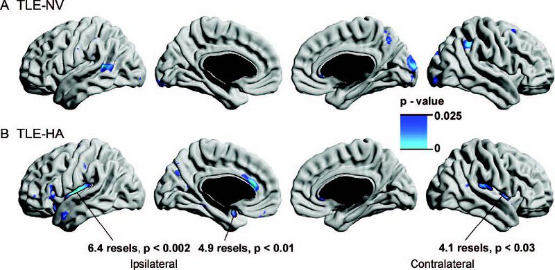
Results: Cross-sectional and longitudinal analyses revealed highly similar topology and rates of neocortical thinning in both TLE groups, predominantly in frontocentral, temporal, and cingulate regions. Bootstrap methods showed that at least 20 subjects per group were necessary to reliably observe these patterns of atrophy in TLE. Moreover, power analysis showed that even with sample sizes of 80 subjects per group, differences in thickness between TLE-HA and TLE-NV would be marginal. With respect to postsurgical outcome, we found an association between residual seizures and atrophy in temporopolar and insular cortices in TLE-HA, and in the posterior quadrant in TLE-NV.
Conclusion: We demonstrated with a high degree of confidence that static and dynamic effects of epilepsy impact similarly the neocortex of patients with hippocampal atrophy and patients with normal hippocampal volumes. On the contrary, areas predicting unfavorable postsurgical outcome were distinct, suggesting different configurations of epileptogenic networks in these 2 groups.