Abstract
Background: Whether recurrent epileptic seizures induce brain damage is debated. Disease progression in epilepsy has been evaluated only in a few community-based studies involving patients with seizures well controlled by medication. These studies concluded that epilepsy does not inevitably lead to global cerebral damage.
Objective: To track the progression of neocortical atrophy in pharmacoresistant temporal lobe epilepsy (TLE) using longitudinal and cross-sectional designs.
Methods: Using a fully automated measure of cortical thickness on MRI, we studied a homogeneous sample of patients with pharmacoresistant TLE. In the longitudinal analysis (n = 18), fixed-effect models were used to quantify cortical atrophy over a mean interscan interval of 2.5 years (range = 7 to 90 months). In the cross-sectional analysis (n = 121), we correlated epilepsy duration and thickness. To dissociate normal aging from pathologic progression, we compared aging effects in TLE to healthy controls.
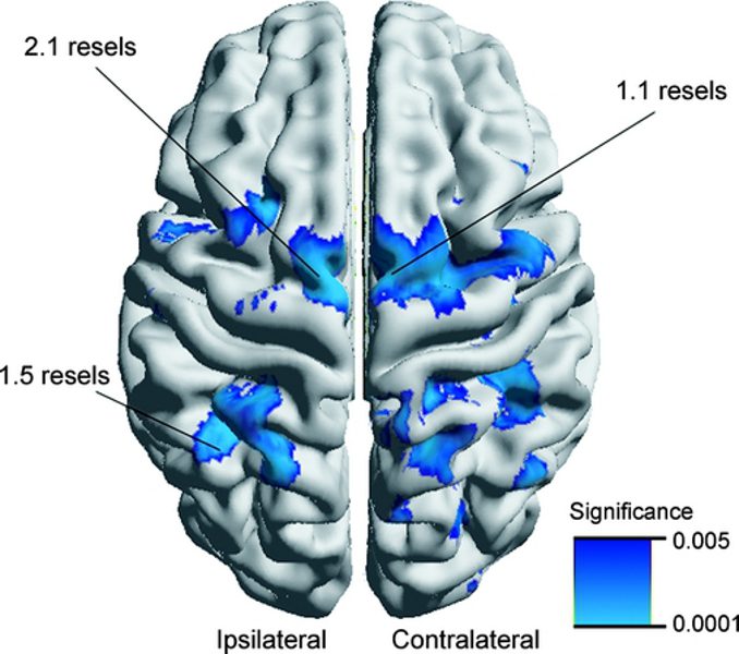
Results: The longitudinal analysis mapped progression in ipsilateral temporopolar and central and contralateral orbitofrontal, insular, and angular regions. In patients with more than 14 years of disease, atrophy progressed more rapidly in frontocentral and parietal regions that in those with shorter duration. The cross-sectional study showed progressive atrophy in the mesial and superolateral frontal, and parietal cortices.
Conclusion: Our combined cross-sectional and longitudinal analysis in patients with pharmacoresistant temporal lobe epilepsy demonstrated progressive neocortical atrophy over a mean interval of 2.5 years that is distinct from normal aging, likely representing seizure-induced damage. The cumulative character of atrophy underlies the importance of early surgical treatment in this group of patients.