Abstract
BACKGROUND & OBJECTIVES: MRI fails to reveal hippocampal pathology in 30% to 50% of temporal lobe epilepsy (TLE) surgical candidates. To address this clinical challenge, we developed an automated MRI-based classifier that lateralizes the side of covert hippocampal pathology in TLE.
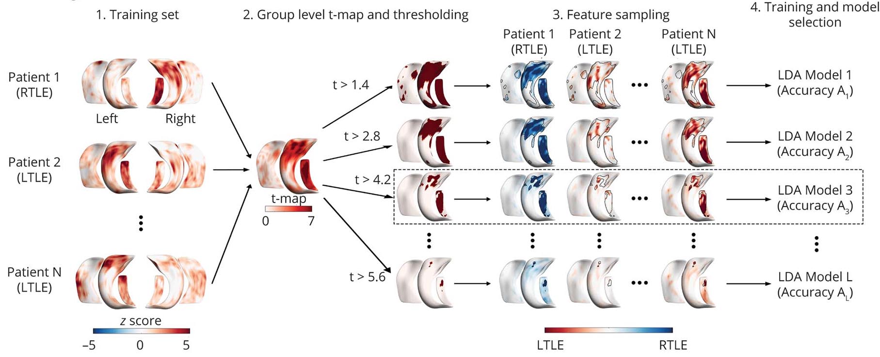
METHODS: We trained a surface-based linear discriminant classifier that uses T1-weighted (morphology) and T2-weighted and fluid-attenuated inversion recovery (FLAIR)/T1 (intensity) features. The classifier was trained on 60 patients with TLE (mean age 35.6 years, 58% female) with histologically verified hippocampal sclerosis (HS). Images were deemed to be MRI negative in 42% of cases on the basis of neuroradiologic reading (40% based on hippocampal volumetry). The predictive model automatically labeled patients as having left or right TLE. Lateralization accuracy was compared to electroclinical data, including side of surgery. Accuracy of the classifier was further assessed in 2 independent TLE cohorts with similar demographics and electroclinical characteristics (n = 57, 58% MRI negative).
RESULTS: The overall lateralization accuracy was 93% (95% confidence interval 92%–94%), regardless of HS visibility. In MRI-negative TLE, the combination of T2 and FLAIR/T1 intensities provided the highest accuracy in both the training (84%, area under the curve [AUC] 0.95 ± 0.02) and validation (cohort 1 90%, AUC 0.99; cohort 2 76%, AUC 0.94) cohorts.
DISCUSSION: This prediction model for TLE lateralization operates on readily available conventional MRI contrasts and offers gain in accuracy over visual radiologic assessment. The combined contribution of decreased T1- and increased T2-weighted intensities makes the synthetic FLAIR/T1 contrast particularly effective in MRI-negative HS, setting the basis for broad clinical translation.
CLASSIFICATION OF EVIDENCE: This study provides Class II evidence that in people with TLE and MRI-negative HS, an automated MRI-based classifier accurately determines the side of pathology.