Abstract
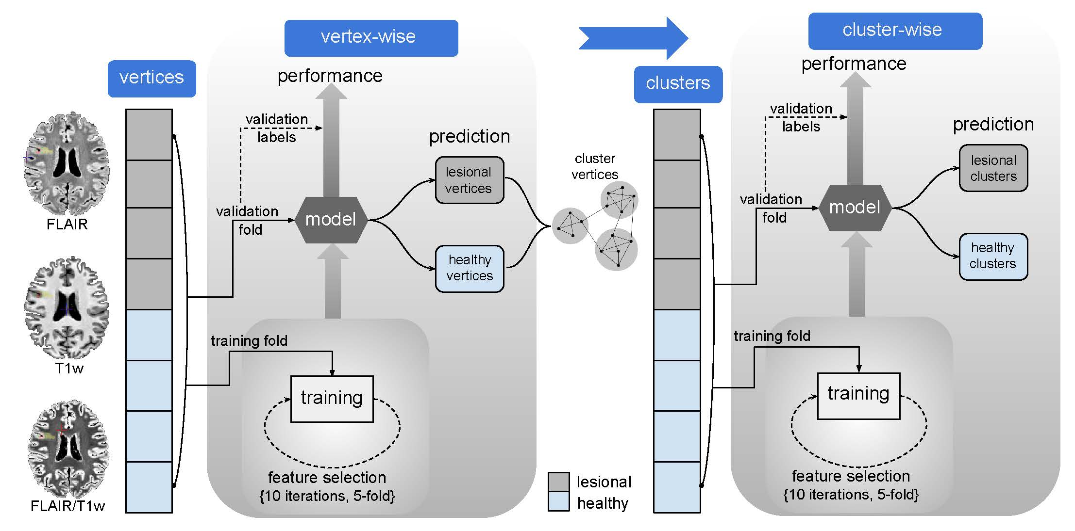
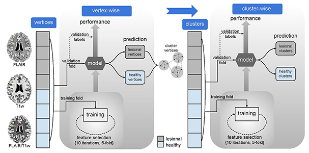
Focal cortical dysplasia (FCD), a malformation of cortical development, is a frequent cause of drug-resistant epilepsy. This surgically-amenable lesion is histologically characterized by cortical dyslamination, dysmorphic neurons, and balloon cells, which may extend into the immediate subcortical white matter. On MRI, FCD is typically associated with cortical thickening, blurring of the cortical boundary, and intensity anomalies. Notably, even histologically-verified FCD may not be clearly visible on preoperative MRI. We propose a novel FCD detection algorithm, which aggregates surface-based descriptors of morphology and intensity derived from T1-weighted (T1w) MRI, T2-weighted fluid attenuation inversion recovery (FLAIR) MRI, and FLAIR/T1w ratio images. Features were systematically sampled at multiple intracortical/subcortical levels and fed into a two-stage classifier for automated lesion detection based on ensemble learning. Using 5-fold cross-validation, we evaluated the approach in 41 patients with histologically-verified FCD and 38 age-and sex-matched healthy controls. Our approach showed excellent sensitivity (83%, 34/41 lesions detected) and specificity (92%, no findings in 35/38 controls), suggesting benefits for presurgical diagnostics.