Resources Available
MR Imaging Facilities
fMRI studies are conducted on the Centre's
1.5 Tesla Siemens Vision scanner, using gradient echo EPI sequences. The
scanner is operated by trained technicians from the radiology department
during fMRI experiments, using preset protocols set up by BIC personnel.
Some flexibility in these protocols is allowed, and you may wish to consult
us on protocol decisions when planning a study.
Stimulus Presentation and Subject Monitoring
Equipment is in place for communicating with
subjects in the scanner via intercom and for presenting visual stimuli
or prompts using an LCD projector (NEC MultiSync MT 1030+, specifications
about the projector can be found at : http://www.nectech.com/presentationproducts/index.htm)
and mirror system. The projector runs at 1024 x 768, and can be connected
to a PC, Mac, SGI, or other computer using a normal DB15 VGA cable (Macs
require an adaptor which we have).
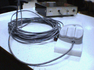
There is also an MR compatible PS/2
mouse available.

MR Compatible Mouse and Synchronization Box
We are currently able to monitor subject feedback
and provide synchronization between data acquisition and stimulus delivery
software by way of a PS/2 mouse emulator. This device can be plugged into
a PS/2 mouse port on a PC or SGI O2 computer (or anything else that takes
a PS/2 mouse) and produces a single middle mouse button click (button down
followed by button up 100 ms later) every time a scan is performed. Subject
feedback from a two button MR compatible mouse
is communicated as left or right mouse button clicks. Note that this device
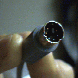
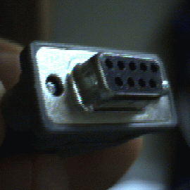
could also be plugged into a serial port. Here are pictures of the mouse
connector we use (and a serial port adaptor), in case you're wondering
if it's compatible with your connectors:


PS/2 Mouse Connector and Serial Adaptor
Some software for the presentation of stimuli/prompts
in synchrony with fMRI data acquisition exists, but this is generally the
responsibility of the experimenter. Timing of the experiment can be controlled
by counting middle button clicks from the PS/2 mouse emulator, and subject
feedback can be recorded by monitoring the left and right mouse buttons.
There is a PC486 in the console room which is used under Linux to telnet
to other machines (like an O2 in the equipment room beside the penetration
panel running stimulation software) and which can also run DOS programs.
If you need anything more you will have to provide it yourself at this
point.
A variety of implements for subject immobilization
are available, including a vacuum head cushion and a bite bar assembly.
The bite bar and mirror assembly are apparatus Dr. R. Hoge built for his
experiments. Because they are used by almost everybody who's doing fMRI
experiments, please do not disassemble them, or if you do, please ensemble
them back the way they were. The apparatus is of modular design and if
parts of it are lost or broken, please let us know, so we can replace them
before the next fMRI study.
Computing Resources for Data Analysis
Following an experiment, the raw data are
transferred from the MR scanner computer and analyzed on the BIC computers
using software tools described below. The technicians perform the data
transfer, and you are responsible for telling them which node to transfer
to (usually the fMRI' node). fMRI studies consume large amounts of disk
space. Because the disks on the scanner get filled quite quickly,
you are also responsible for notifying the techs as soon as you have verified
that the transfer was successful. It is recommended that you verify this
as
early as possible, because sometimes the transfers are completely
or partially unsuccessful and people have lost entire sessions. The
techs will delete the data after you notified them, unless you had transfer
problems and they will transfer your data again. If you don't notify
the techs, data will stay on the scanner disks for
24 hours, after
this period, the data will be deleted.
We'd like to provide
backup of the raw fMRI data on the the scanner, but this is not currently
feasible.
All data backup is therefore the responsibility of the
experimenter.
The experimenter is responsible for data
analysis, and one of the main purposes of this document is to show those
conducting fMRI studies how to use the software available to analyze their
data.
The following sections walk you through
the steps required to plan a study, book the scanner, and do the experiments
and analyze and interpret the data.
Planning The Study
Approval
fMRI studies at the Neuro have to be approved
by the MNI MR Research Committee and the MNI Research Ethics Committee.
Stimulation and Feedback
You will need to work out how you are going
to present stimuli or prompts to subjects in the MR scanner, as well as
record any feedback that you require. Some equipment and software is in
place for this purpose, but meeting specialized needs is the responsibility
of the experimenter. If you plan to use your own computer for your study,
there are two important facts you should take into account:
The LCD projector used to show visual
stimuli to the subject runs in 1024 x 768 mode and below. Also,
if you are going to use the head coil for the experiment, it is not possible
to see the entire screen area with the current coil and display setup.
You should try out the display and take this into account when setting
up the experiment.
Bringing a color monitor into the MRI suite
may permanently damage it, so the software for your experiment should not
require a color CRT for controlling the experiment from the console room.
Some people have used old color monitors.
Task Design
Subject motion must be kept to an absolute
minimum, and this fact must be kept in mind when designing experimental
paradigms.
Because of the stringent immobilization
requirements, subjects generally can not tolerate scanning sessions which
last longer than about 1.5 hours.
Booking Scanner Time
Scanner time on the Siemens Vision system
can be booked through the receptionist (ext. 8510) at the MRI front desk
on 3B. Make sure you book enough time to carry out all the experiments
you are planning, including setup time and possible delays between scanning
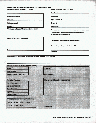
runs. Before you can book scanner time, make sure you have a consent
form and a requisition form, filled in and signed.
The requisition form looks like the form
in the picture below:

Please make sure you fill in the Ethics Approval # and the Account #.
SAFETY ISSUES
There are a number of safety issues to keep
in mind during an MRI study. These involve various dangers posed by the
strong magnetic field that exists in the scanning room.
NO FERROMAGNETIC METAL OBJECTS IN THE MR SCANNER
ROOM!
These are attracted to the magnet with great
force, and can pose an extreme hazard. If you are in doubt about an object,
ask the techs or physics personnel
before bringing it into the room.
Examples of potentially dangerous objects include the following:
scissors
screwdrivers and wrenches
coins
pencils and pens with metal parts
any metallic implant in a patient, subject,
or investigator (ask the techs if you're not sure)
Before you enter the scanner room,
please make sure that your subject /patient doesn't have metallic clips,
metallic implants or a pacemaker, or any other ferromagnetic metal objects.
Things which are metal but may go into
the
room (but not necessarily the scanner) include
zippers and rivets on clothing (jeans
are ok)
metal eyelets on footwear
belt buckles (still better to remove from
subject or patient)
most eyeglass frames (if in doubt, hold
onto glasses when entering room - metal frames must never be used
by subjects inside the scanner, as they may pose a current loop hazard)
NO POTENTIAL CURRENT LOOPS INSIDE MAGNET
Where electrically conducting materials such
as wires must be placed inside the magnet bore (e.g. EEG, EOG, stimulation
devices), care must be taken to avoid conducting loops. The system can
induce strong currents in these, and burns to the subject may result. Consult
physics personnel if in doubt.
The scanner will wipe out magnetic strip
cards (credit cards, bank cards, etc.) and can damage watches. These
should be left in the console room.
The scanner may damage computer monitors.
Any CRT display will be distorted if brought near the MR suites. Color
CRTs in particular may be permanently damaged. LCD displays, such as those
used on notebook computers, are not affected.
Carrying Out the Experiments
The usual sequence of events is as follows:
You show up at the MRI unit with your
subject. The MR Research committee has approved your study and protocol,
and the subject has signed your MNI REC-approved consent form.
Individuals who will enter the room
containing the MRI scanner remove all metal objects, magnetic cards, and
watches.
You go over the protocol with the techs
and set up any stimulus/prompting/feedback equipment and software you need.
The subject is placed in the scanner by
the techs and the immobilization system is adjusted.
A series of anatomic scans is performed:
A 30 second scout acquisition
A 10-14 minute T1-weighted 3D anatomic
scan
The functional scanning runs are performed.
Each usually lasts about six minutes, but this depends on the number of
frames you have and the repetition time between frames. If you are using
the high resolution protocols (128 x 128), there may be long (up to five
minutes) delays between scans while the system digests data acquired in
the previous run. This should be taken into account when estimating the
amount of time required to carry out a given number of scans. The delays
get longer the more slices are acquired.
Subject gets removed from scanner.
You recover any equipment you set up in
the MRI suite (and don't forget your watches and wallets!)
You tell the technician where to transfer
the data (usually the fMRI node, which results in the scans appearing in
~fmrixfer/images on bottom).
The transfer, which may take several hours,
is initiated.
Be sure to verify that all scans have
been successfully transferred, then let the techs know that it is ok for
them to delete your study. Data on the scanner will be deleted after
24 hours unless you notify the technicians of any problems (in person or
at ext. 8510).


