Abstract
Objective: Although experimental work has provided evidence that the thalamus is a crucial relay structure in temporal lobe epilepsy (TLE), the relation of the thalamus to neocortical pathology remains unclear. To assess thalamocortical network pathology in TLE, we mapped pointwise patterns of thalamic atrophy and statistically related them to neocortical thinning.
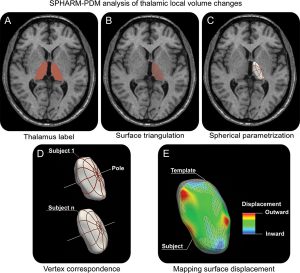
Methods: We studied cross-sectionally 36 patients with drug-resistant TLE and 19 age- and sex-matched healthy control subjects using high-resolution MRI. To localize thalamic pathology, we converted manual labels into surface meshes using the spherical harmonic description and calculated local deformations relative to a template. In addition, we measured cortical thickness by means of the constrained Laplacian anatomic segmentation using proximity algorithm.
Results: Compared with control subjects, patients with TLE showed ipsilateral thalamic atrophy that was located along the medial surface, encompassing anterior, medial, and posterior divisions. Unbiased analysis correlating the degree of medial thalamic atrophy with cortical thickness measurements mapped bilateral frontocentral, lateral temporal, and mesiotemporal cortices. These areas overlapped with those of cortical thinning found when patients were compared with control subjects. Thalamic atrophy intensified with a longer duration of epilepsy and was more severe in patients with a history of febrile convulsions.
Conclusion: The degree and distribution of thalamic pathology relates to the topography and extent of neocortical atrophy, lending support to the concept that the thalamus is an important hub in the pathologic network of TLE.
