Abstract
Background: We previously demonstrated that shape variants of the hippocampal formation are more prevalent in patients with temporal lobe epilepsy (TLE) than in healthy individuals.
Objective: To categorize sulcal patterns of the basal temporal lobe in TLE compared to healthy controls.
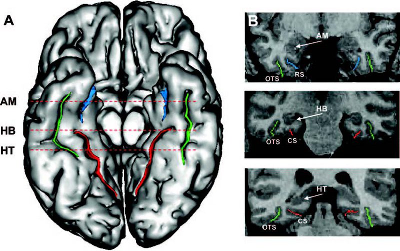
Methods: We studied 51 healthy controls and 69 patients with TLE (37 left, 32 right TLE). Brain sulci were identified and labeled automatically on MRI using an algorithm based on a congregation of neural networks that allows mapping three-dimensional sulcal models on the cortical surface. We used four sulcal patterns classes to categorize the sulcal arrangement in the inferior surface of the temporal lobe in each subject: Type 1, i.e., single-branch, unbroken collateral sulcus (CS) connected with the rhinal sulcus (RS) anteriorly; Type 2, i.e., CS connected with the occipitotemporal sulcus (OTS), but separated from the RS; Type 3, i.e., CS separated from the OTS and RS, which are connected; and Type 4, i.e., CS, OTS and RS separated.
Results: In healthy controls, Type 1 and Type 2 were the patterns seen most frequently. Overall, 82% (42/51) of subjects had the same sulcal pattern in both temporal lobes. Inter-rater reliability for 35 randomly selected subjects indicated excellent agreement (Cohen's Kappa: 0.84). Compared to controls, we found an increased frequency of Type 1 CS in patients with TLE, both in the left (77% vs 47%, p = 0.004) and the right hemispheres (72% vs 41%, p = 0.002). On the other hand, we found a decreased frequency of Type 2 CS in patients with TLE, both in the left (4% vs 31%, p = 0.00002) and the right hemisphere (4% vs 35%, p < 0.00001).
Conclusion: A single-branch, unbroken collateral sulcus is the predominant sulcal pattern found in temporal lobe epilepsy. This "simplified" arrangement may be an indicator of neurodevelopmental deviance associated with this condition.