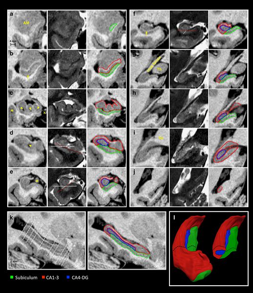
Abstract
The hippocampus is composed of distinct anatomical subregions that participate in multiple cognitive processes and are differentially affected in prevalent neurological and psychiatric conditions. Advances in high-field MRI allow for the non-invasive identification of hippocampal substructure. These approaches, however, demand time-consuming manual segmentation that relies heavily on anatomical expertise. Here, we share manual labels and associated high-resolution MRI data (MNI-HISUB25; submillimetric T1- and T2-weighted images, detailed sequence information, and stereotaxic probabilistic anatomical maps) based on 25 healthy subjects. Data were acquired on a widely available 3 Tesla MRI system using a 32 phased-array head coil. The protocol divided the hippocampal formation into three subregions: subicular complex, merged Cornu Ammonis 1, 2 and 3 (CA1-3) subfields, and CA4-dentate gyrus (CA4-DG). Segmentation was guided by consistent intensity and morphology characteristics of the densely myelinated molecular layer together with few geometry-based boundaries flexible to overall mesiotemporal anatomy, and achieved excellent intra-/inter-rater reliability (Dice index ≥90/87%). The dataset can inform neuroimaging assessments of the mesiotemporal lobe and help to develop segmentation algorithms relevant for basic and clinical neurosciences.