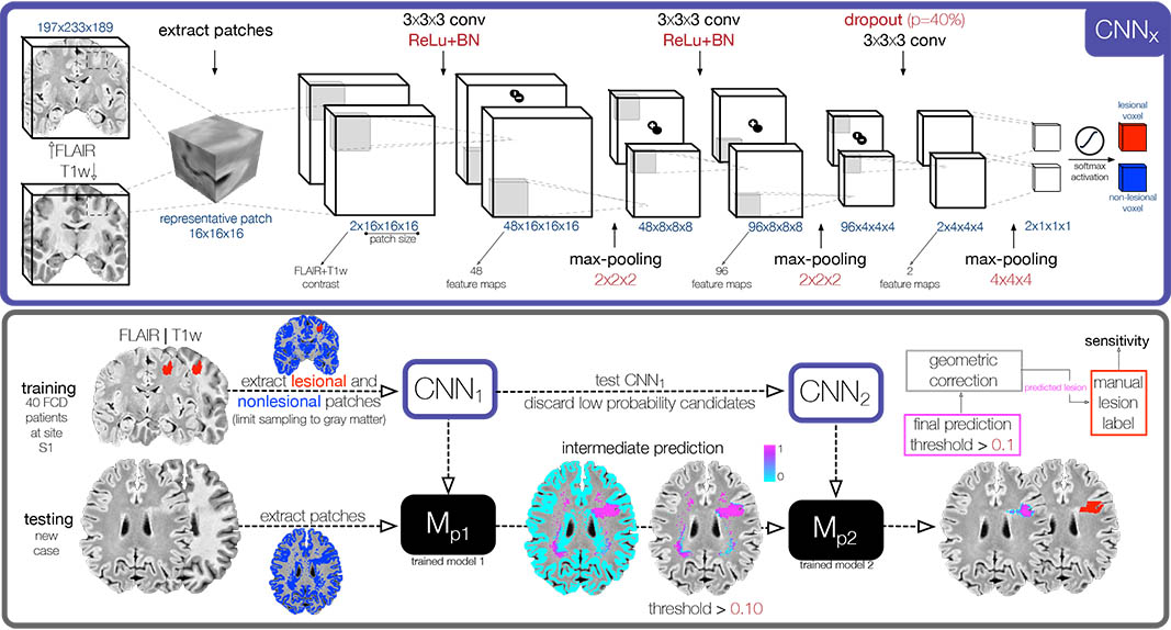
Abstract
Focal cortical dysplasia (FCD) is a prevalent surgically-amenable epileptogenic malformation of cortical development. On MRI, FCD typically presents with cortical thickening, hyperintensity, and blurring of the gray-white matter interface. These changes may be visible to the naked eye, or subtle and be easily overlooked. Despite advances in MRI analytics, current surface-based algorithms fail to detect FCD in 50% of cases. Moreover, arduous data pre-processing and specialized expertise preclude widespread use. Here we propose a novel algorithm that harnesses feature-learning capability of convolutional neural networks (CNNs) with minimal data pre-processing. Our classifier, trained on a patch-based augmented dataset derived from patients with histologically-validated FCD operates directly on MRI voxels to distinguish the lesion from healthy tissue. The algorithm was trained and cross-validated on multimodal MRI data from a single site (S1) and evaluated on independent data from S1 and six other sites worldwide (S2–S7; 3 scanner manufacturers and 2 field strengths) for a total of 107 subjects. The classifier showed excellent sensitivity (S1: 87%, 35/40 lesions detected; S2–S7: 91%, 61/67 lesions detected) and specificity (S1: 95%, no findings in 36/38 healthy controls; 90%, no findings in 57/63 disease controls). Easy implementation, minimal pre-processing, high performance and generalizability make this classifier an ideal platform for large-scale clinical use, particularly in "MRI-negative" FCD.