Abstract
Objective: Converging evidence suggests that abnormalities of brain development may play a role in the pathogenesis of temporal lobe epilepsy (TLE). As sulco-gyral patterns are thought to be a footprint of cortical development, we set out to quantitatively map folding complexity across the neocortex in TLE. Additionally, we tested whether there was a relationship between cortical complexity and features of hippocampal maldevelopment, commonly referred to as malrotation.
Methods: To quantify folding complexity, we obtained whole-brain surface-based measures of absolute mean cortical curvature from MRI scans acquired in 43 drug-resistant patients with TLE with unilateral hippocampal atrophy, and 40 age- and sex-matched healthy controls. In patients, we correlated changes in cortical curvature with 3-dimensional measures of hippocampal positioning.
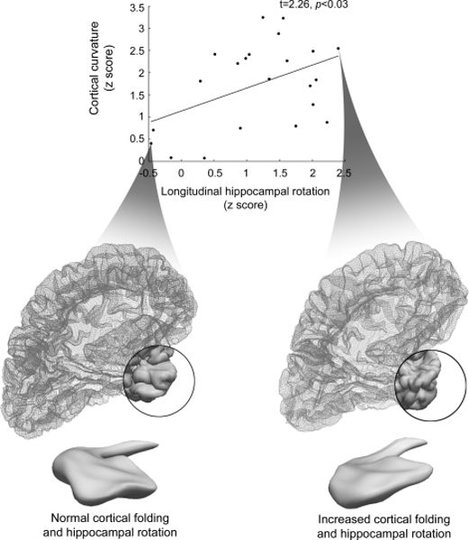
Results: We found increased folding complexity in the temporolimbic cortices encompassing parahippocampal, temporopolar, insular, and fronto-opercular regions. Increased complexity was observed ipsilateral to the seizure focus in patients with left TLE (LTLE), whereas these changes were bilateral in patients with right TLE (RTLE). In both TLE groups, increased temporolimbic complexity was associated with increased hippocampal malrotation. We found tendencies for increased complexity in bilateral posterior temporal cortices in LTLE and contralateral parahippocampal cortices in RTLE to be predictive of unfavorable seizure outcome after surgery.
Conclusion: The anatomic distribution of increased cortical complexity overlapping with limbic seizure networks in TLE and its association with hippocampal maldevelopment further imply that neurodevelopmental factors may play a role in the epileptogenic process of TLE.