Images Obtained from the Processing of the MNI Phantom (Noise 0%; RF 0%;) avalaible at http://www.bic.mni.mcgill.ca/brainweb
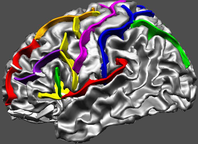
Image One : Inner Cortical Surface/Sulci
A 3-D rendering of automatically extracted and identified cortical sulci superimposed on the inner cortical surface (i.e. the grey matter/white matter interface). Such modeling of cortical structures, obtained from 3D MRI, can be used in several contexts including surgical planning, interpretation of functional examinations (MEG, PET, fMRI,...), extraction of statistical information representing normal and/or pathological variability of the cortical topography.
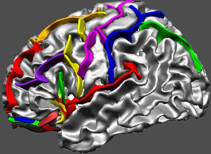
Image Two: White Matter Surface and Sulcus Superficial Parts
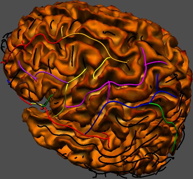
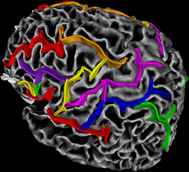
Image Three: Sulcus Extraction and Automatic Labelling
( 8 Main Sulci). See table for colours legend.
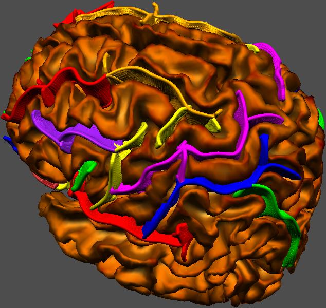
Image Four : Corresponding Manual Labelling
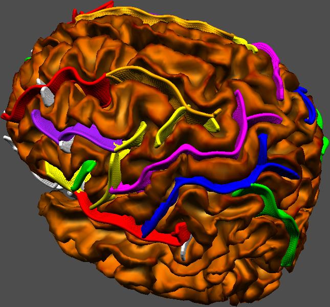
Image Two and Three : Left (or Top) Manual Labelling; Right (or Bottom) Automatic Labelling. (see Table color legend).