Most non-linear registration algorithms compute a continuous spatial mapping from the coordinate system of a given subject's MRI to that of a target MRI volume. It is assumed that this mapping incorporates all morphological differences between subject and target anatomies. We turn this paradigm around to simulate different anatomies, based on a single target volume. Random spatial deformations were generated and applied to the brain phantom to build a family of homologous, but morphologically different brain phantoms. An MRI volume was simulated for each member of the family.
Random spatial deformations were generated by defining a set of twenty
landmarks within the target brain volume. A second deformed set of
landmarks was produced by adding a random displacement to each of the
landmark coordinates. Since previous work in our laboratory has shown
anatomical variability to be on the order of 4-7mm [25], a
Gaussian random number generator with a standard deviation of 5mm was
used to produce each component of the displacement vector. The two
resulting point sets (original and deformed) were then used to define a
continuous 3-D thin-plate spline transformation function. Ten such
deformations were generated and used to resample the original target data
and produce 10 spatially warped source data sets for testing. The average
deformation magnitude was 7.7mm with a maximum of 19.7mm. Figure
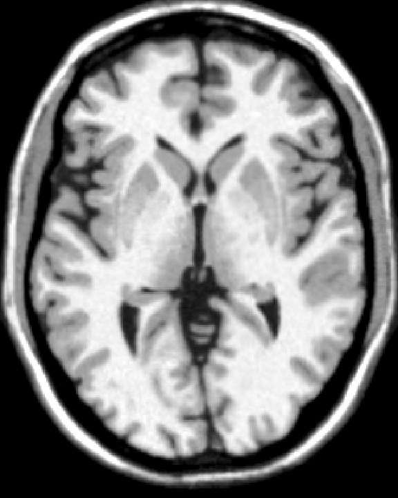
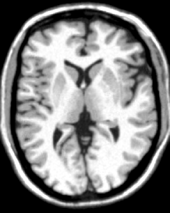
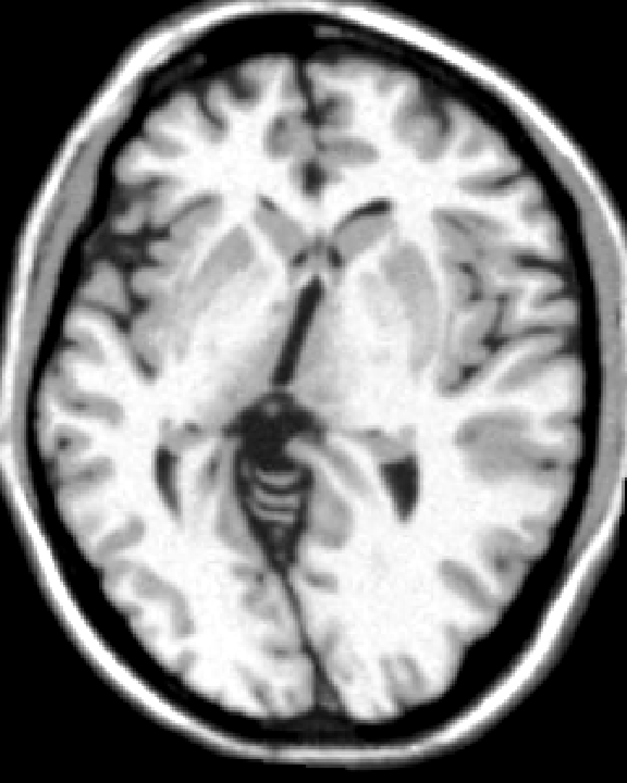
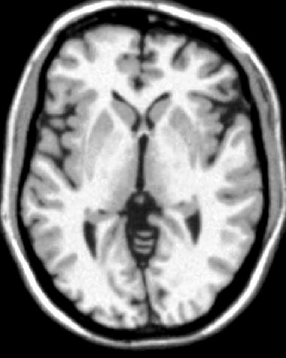
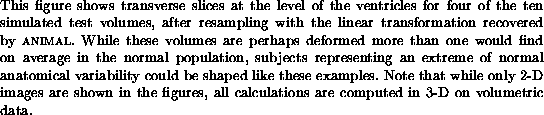
1 shows transverse slices (after linear stereotaxic
registration) through the original and three of the ten warped volumes used
in the experiments below. While these images demonstrate extreme
deformations, they form a good test for
![]() .
.