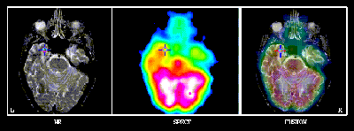
In using the internal landmark matching technique for registering brain volumes from single photon emission computed tomography (SPECT) and magnetic resonance (MR) imaging, the resulting errors are different from similar registrations of positron emission tomography (PET) and MR volumes because of the anisotropic nature of SPECT's tomographic resolution. SPECT/MR registration errors are investigated with point simulations to allow a controlled study of the dependence of the translation and rotation errors on different factors. These factors include: the number of point pairs, m, the error in point pair homology (characterised by the FWHM of a 3-D Gaussian error envelope), and the configuration of the point sets (as they depend on their radius from their centroid). The results of this study are used to interpret the results from studies based on a 3-D brain phantom fitted with external fiducials to provide the basis for true registrations.
It is found that the same error distributions may not be expected across
ordinates. Based on the imaging capabilities of the sensors used in this
study, (a) maximum translation errors of mm in the
anterior-posterior orientation are found to be a consequence of SPECT's
anisotropic resolution and the brain's major axis in that orientation, and
(b) rotation errors of about
in
the coronal plane are larger than in the transaxial because of
the comparative dimensions in those planes.
This work would not have been possible without the aid of numerous people. Primarily, I am extremely humble and grateful to my parents and family for their inveterate support and care throughout my academic vacation. Not less importantly, I would like to express my admiration and gratitude to my supervisors, Dr. Geoffrey W. Dean and Dr. Alan C. Evans, for their wisdom, guidance and inspiration. This study would not have been possible without them. Peter Neelin, Lise Proulx, Pierre Prévost, Paule Toussaint, Dr. Cécile Sorlié and the staff of the Department of Nuclear Medicine at the Royal Victoria Hospital (RVH, where the SPECT scans were acquired) and the Positron Imaging Lab at the Montreal Neurological Institute (MNI, where much of the computation was performed) were true gems for their amicability and wealth of irreplaceable practical experience. Ian Brown, Nick Waterton, Marc St. Laurent, and Richard Mercier of GE Canada were all always receptive to academic research and aided tremendously with the SPECT systems. Appropriate thanks must also be extended to Vasco Kollokian for his general patience and in appreciation of his network administration talents. Much of the clinical insight into the implications of the study was afforded by Dr. Robert Lisbona (RVH), Dr. François Dubeau (MNI), Dr. Deirdre McMackin (MNI), &Dr. David C. Reutens (MNI). I would like to thank all of them for their advice, enlightenment and humor. I would especially like to offer my most sincerest sentiments of respect and humility to the faculty, staff and students (past &present) of the Medical Physics Unit in light of their collective work in establishing the much respected reputation of the MPU. Their efforts have allowed me to ride on particularly distinguished coat tails.
A. M. D. G.
-(AjfL)-