Stereotaxy is the methodology involved in the three-dimensional localization of structures within the brain, based on diagnostic image information, and the use of surgical instruments to reach these points. Traditionally, stereotaxy has been used to:
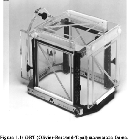
Conventional stereotaxy makes use of a frame attached to the patient's head. The OBT (Olivier-Bertrand-Tipal) frame, shown in OBT_frame, is currently used at the Montreal Neurological Institute both for relating the patient to the image coordinate systems, as well as a support for surgical instruments in the operating room (OR).

Horsley and Clarke, in 1908, described the first stereotactic procedure on small animals [29]. They used a frame, fixed with respect to external anatomical landmarks, to place electrodes at specific points in the animal's brain. Because this technique was based on the false assumption that the position of an internal point was determined solely by the position of external landmarks, it was not precise enough to be used on human subjects.
The next major step in stereotaxy occured in 1947, when Spiegel and Wycis used an imaging technique called pneumoencephalography (which consists in x-ray images of the brain with air injected into the ventricular space) [58]. This provided information about spatial localization of soft tissue landmarks (for example the pineal gland) with respect to the stereotactic frame. Since the specific anatomy of each subject was taken into account, this technique could be used in humans and is the precursor of the first human brain atlas. During this period, Leksell and Riechert [37] in Sweden, and Talairach [61] in France, developed their own stereotactic systems, also based on projection images.
The limitations of all these systems were strongly related to the imaging technique used. Conventional radiographic techniques did not allow unambiguous localization of points in three-dimensional space since they provided only a projection of the subject along one dimension, making the positions of structures perpendicular to the imaging plane indeterminate.
The advent of Computed Tomography (CT) [30] gave stereotaxy a new life, providing three-dimensional representations of the subject. Then, it became possible, in principle, to determine from the image set, the position in three-dimensional space of any point inside the brain. Later, Magnetic Resonance Imaging (MRI) [36,10] provided another alternative for stereotaxy, offering anatomical information complementing that of CT. Moreover, Digital Subtraction Angiography (DSA) [63] and Magnetic Resonance Angiography (MRA) made their appearance, making possible the three-dimensional representation of vasculature. Positron Emission Tomography (PET) has also proved itself useful to provide functional information.
The basis of any stereotactic procedure is the establishment of a reference coordinate system common to both the diagnostic information provided by the images (image space) and the patient's space (surgical space). This relies on the ability to localize the same objects or structures in both spaces, a function that is provided by the stereotactic frame. Typical stereotactic frames use N-shaped markers (for CT or MRI) or point markers (for DSA) that allow the position of structures within a volume to be determined unambiguously [28,20]. Galloway et al. [18] presents a comparison of the accuracy of four different frame-based stereotactic systems.
Until recently, stereotaxy was confined to the pre-surgical planning of procedures. Image-guided neurosurgery (), on the other hand refers to the guidance, on the basis of images, interactively during the procedure. Moreover, allows the guidance in open-cranium surgeries, which is not possible in conventional stereotaxy because of the presence of the frame.