In order to create a 3-D model of the patient's head and brain
surfaces from MRI, the data must first be segmented. In our clinical
environment this is performed on an Allegro workstation,
a product of ISG Technologies![]() ,
where the image is segmented by thresholding its intensity to the
desired value. Whenever this technique fails to give an accurate
contour of the region of interest (due to image artifacts or poor
contrast between the skin and the background), the user can manually
correct the contour. User interaction of this type is also used for
the segmentation of internal brain structures and lesions [62].
,
where the image is segmented by thresholding its intensity to the
desired value. Whenever this technique fails to give an accurate
contour of the region of interest (due to image artifacts or poor
contrast between the skin and the background), the user can manually
correct the contour. User interaction of this type is also used for
the segmentation of internal brain structures and lesions [62].
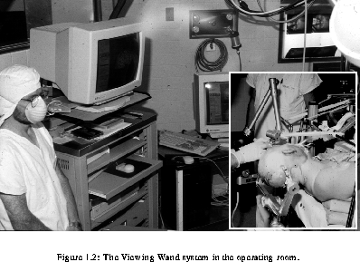
The preparation of a typical procedure involves the following steps. First, a three-dimensional MR or CT volume must be acquired. In some cases, extrinsic fiducial markers are desired to increase the number of visible landmarks, and they must be placed on the patient's head prior to scanning. The next stage involves the transfer of data from the scanner to the Allegro workstation. Then, segmentation of the skin, brain, lesion and other relevant internal structures is performed. This is largely a manual procedure and can take from 20 minutes to two hours, depending on the number and complexity of structures to be segmented and on the number of slices in the data set. The resulting contour data are then processed to form 3-D reconstructions of the surface of the segmented structures. The original acquisition data along with reconstructed three-dimensional objects are then transferred to a second workstation in the operating room.