Two series of three experiments were completed varying only the features
used by ![]() where the first series was completed for simulated data,
and the second series, for real MRI data. In each experiment, 10 MRI
volumes were registered to the chosen target volume. The 3 different
experiments were defined as follows: (1)
where the first series was completed for simulated data,
and the second series, for real MRI data. In each experiment, 10 MRI
volumes were registered to the chosen target volume. The 3 different
experiments were defined as follows: (1) ![]() in standard non-linear
mode, (2)
in standard non-linear
mode, (2) ![]() +selected sulci, (3)
+selected sulci, (3) ![]() +all sulci after using
selected sulci.
+all sulci after using
selected sulci.
In method 2, the problem of establishing correspondence is addressed by
using ten extracted sulci (superior and middle frontal, Sylvian, olfactory
and central, on both hemispheres) as features in addition to the blurred
MRI intensity volume already used by ![]() , The third method is an
attempt to improve the second, by adding all sulci automatically extracted
by
, The third method is an
attempt to improve the second, by adding all sulci automatically extracted
by ![]() into the registration process after completing the
into the registration process after completing the
![]() +selected sulci registration. A linear transformation is also
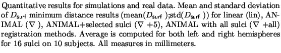
presented for comparisons. Figures 2 and 3
summarize the results qualitatively for simulated and real data,
respectively. Tables 1 and 2 present quantitative results for the measure
described above.
+selected sulci registration. A linear transformation is also
presented for comparisons. Figures 2 and 3
summarize the results qualitatively for simulated and real data,
respectively. Tables 1 and 2 present quantitative results for the measure
described above.
| TABLE 1 | TABLE 2 | |||||||||
| simulations | real MRI | |||||||||
| lin |
|
lin |
|
|||||||
| Central | 3.2;1.5 | 1.2;1.4 | 0.7;0.3 | 0.7;0.4 | 4.7;1.3 | 4.1;1.4 | 2.5;1.2 | 2.5;1.2 | ||
| Postcentral | 4.2;2.3 | 2.2;2.4 | 1.3;1.5 | 1.3;1.5 | 6.4;1.7 | 6.1;2.0 | 5.0;1.7 | 5.0;1.8 | ||
| Middle Frontal | 3.2;1.0 | 1.4;1.2 | 0.7;0.6 | 0.7;0.6 | 6.2;3.5 | 5.4;3.2 | 4.5;3.1 | 4.7;3.2 | ||
| Superior Frontal | 3.4;1.9 | 1.8;2.1 | 1.6;2.1 | 1.7;2.1 | 6.2;3.8 | 5.6;3.6 | 4.4;3.9 | 4.4;4.0 | ||
| Sylvian | 4.0;1.3 | 3.0;1.2 | 2.4;0.9 | 2.4;1.0 | 5.2;1.6 | 5.0;1.4 | 3.6;0.9 | 3.7;1.0 | ||
| Olfactory | 3.2;4.2 | 1.7;2.8 | 1.5;2.9 | 1.6;2.8 | 2.1;1.7 | 1.6;1.9 | 1.3;1.6 | 1.4;1.8 | ||
| average (n=16) | 3.4;2.3 | 2.1;2.1 | 1.6;1.7 | 1.5;1.7 | 5.0;3.2 | 4.5;3.1 | 4.0;3.1 | 4.0;3.1 | ||