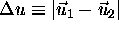
The goal of this study was to determine the size of the registration
error and the variability involved in registrations performed in the
clinical environment (operating room) when procedures were
performed using 3-D images based on MR data. Although these results
have a limited statistical significance and bring few insights to the
mechanism of registration, they demonstrate that the registration
process using MR images is far from trivial, and that the accuracy
currently available using the standard approach is  4 mm at best.
These measurements were performed using the following protocol.
 , the translation vector
  and the scale factor S that constitute the transformation
between the imaging and surgical spaces:
and the scale factor S that constitute the transformation
between the imaging and surgical spaces:
It is not immediately obvious why these graphs are curved lines, since they represent the distance between two straight lines in 3-D space. However, if the lines are represented as:

where  is a scale parameter,
  and
and  are points belonging to the lines,
  and
and  are unit
vectors parallel to them and t is a parameter labelling points on
them, we can compute the difference
are unit
vectors parallel to them and t is a parameter labelling points on
them, we can compute the difference  between corresponding
position t on theses lines:
between corresponding
position t on theses lines:
This will give a ``v-shaped'' function if:

So, the graphs are straight if  is
parallel to
is
parallel to  or if one of these vectors is zero.
In all other cases, the line will be curved.
or if one of these vectors is zero.
In all other cases, the line will be curved.
It can be shown that the curve reaches a minimum value when:

where 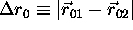 ,
,  and
and  is the angle between
  and
and  .
.
In order that the minimum be at the origin, let  . distance then becomes:
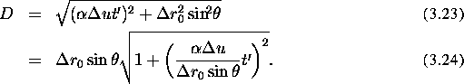
When  ,
,  i.e.,
the distance between the lines increases linearly. When
i.e.,
the distance between the lines increases linearly. When  , we can develop D in a Taylor series:

This shows that the curvature becomes infinite, i.e., the graph is
v-shaped, if  or
or  . If
. If  , i.e., there is no rotational error and
  is constant.
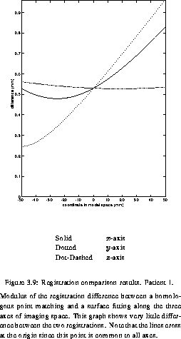
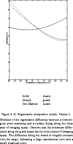
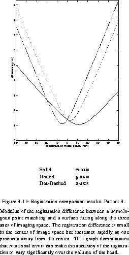
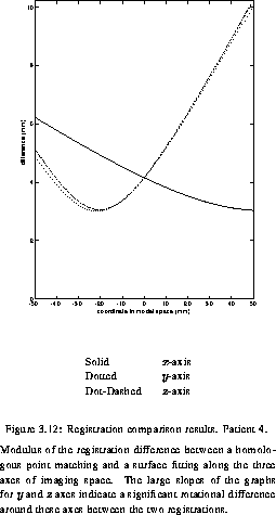
first_patient shows a sub millimetric difference between the two registrations over the volume of 1000 cubic mm. The situation is quite different for the three other patients, where large differences can be observed. The graph for Patient 2 (second_patient), reveals a difference at the origin of 6 mm. This means that, if the surgeon holds the probe at the point corresponding the the origin of the scanner space (roughly in the center of the head), the position of the probe displayed on the screen would differ by 6 mm for the two registrations. Moreover, we can see that the minimum difference is around -25 mm along the x-axis and around +40 mm along the two other axes. This illustrates the fact that even though the registration error is likely to be minimum at the origin, it is not always the case. third_patient shows an example where the rotational error makes the difference between the two registrations increase rapidly, demonstrating that the registration error can be dramatically space-variant. Since the experimental protocol was consistent across the four patients, we believe that the discrepancy observed between the first patient and the other three is due to statistical fluctuation.
As will be demonstrated in Chapter 5, the image of the corners of the frame markers can be subject to displacements from their real position by approximately 2 mm on average. This, coupled with the imprecision due to the fact that it is not possible to physically reach the intersection points of the markers, makes the uncertainty on the point position to be approximately 3 mm. The observation of simul_4_3 shows that the observed errors are certainly possible, although a rigorous comparison is not possible since the curve in simul_4_3 and the curves in figures first_patientlast_patient do not represent the same thing (the latter result represents differences between two different registration techniques while the first is the expected difference between different registrations performed with the same registration technique).

