At our institution cerebral angiograms are routinely acquired stereoscopically. Stereo views are obtained by using a C-arm based digital subtraction angiographic system and performing the angiographic sequence twice at gantry angulations separated by approximately seven degrees. We also use a system, equipped with a stereoscopic (dual focus) tube, that allows acquisition of both stereoscopic DSA and fluoroscopy.
In order to manipulate targets in 3-D space, we must establish the
relationship between points seen in the images and the corresponding
points in the 3-D volume. In the case of frame-based stereotaxy, we
are able to use the fiducial markers, situated at specific 3-D locations,
for this task. The fiducial marker system for DSA consists of pairs of
plates located on the front, back and sides of the frame. Four steel
pellets (0.5 mm diameter) are embedded in each plate at the vertices
of a 70mm square. For a given projection, eight markers appear in the
image: four from the plate closest to the x-ray tube and four from the
plate on the opposite side. Registration is accomplished by first having the
surgeon point to each frame-based fiducial using the probe, and then
computing the rigid-body transformations that relate the two systems.
Full details of this registration approach are given by Henri
et. al.[4]. Once registration is achieved, the stereoscopic system is
operated in a mode that simply tracks the position and orientation of
the probe, then transforms coordinates from the probe to the frame and
displays the probe as it relates to the angiogram floating in
(stereoscopic) space. We have demonstrated elsewhere that a 3-D
localization accuracy of 1mm may be achieved using both AP and
lateral DSA images, or
1mm in plane and
4-5mm in depth using
a single stereoscopic pair[4]. An example of a stereo pair
of DSA images is shown in Fig. 5.
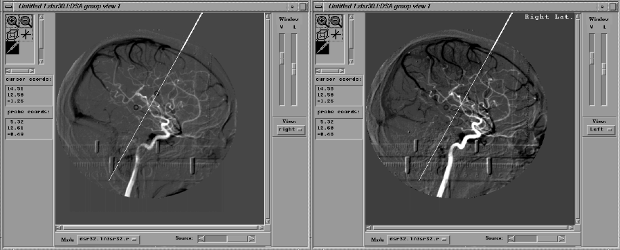 Figure 5: Click on image.