Although DSA provides accurate localization of vascular structures it is not appropriate in all situations due to the unavoidable radiation exposure of the patient and the need for the stereotactic head frame. MRA[18] provides details of the vasculature without these limitations although at lower resolution and accuracy. Unlike DSA which can be displayed only in the plane in which the projections were recorded, MRA has the additional advantage that it can be viewed from an arbitrary 3-D orientation allowing significantly more flexibility for the neurosurgeons when visualizing intracranial trajectories. Additionally, we have found stereoscopic display of MRA data removes ambiguity regarding the relative location of the vessels and the hand-held probe.
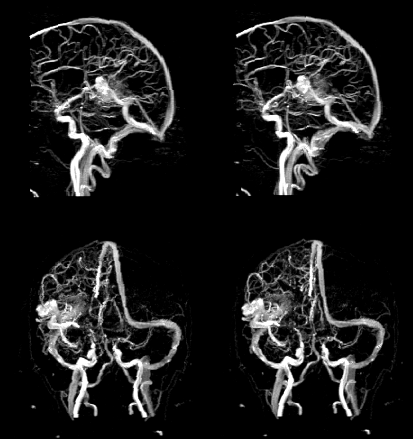
We find both surface-rendered and volume-rendered representations of the MRA data to be useful. Surface-rendering techniques require that the original slice data be segmented to create a 3-D object comprising the vasculature, a task that requires considerable effort with our current segmentation tools. Thus, we usually apply volume rendering techniques, in this case maximum intensity projection (MIP), to the acquired data directly thereby avoiding the segmentation step. Two stereo pairs of MRA images of a patient exhibiting an AVM are displayed in Fig. 6, one from a lateral orientation and one from a more frontal orientation. Eac pair of images was obtained by creating MIP renderings of the 3-D data set from viewpoints differing by 5 degrees.
 Click on image.
Click here for movie.
Click on image.
Click here for movie.