A set of three spin-echo acquisitions was obtained. One of these was a
standard sequence, whereas the other two were performed with the
 pulse shifted in time by
  1.12 ms. The phase and
magnitude images of the three acquisition are shown in
se_acq.
1.12 ms. The phase and
magnitude images of the three acquisition are shown in
se_acq.
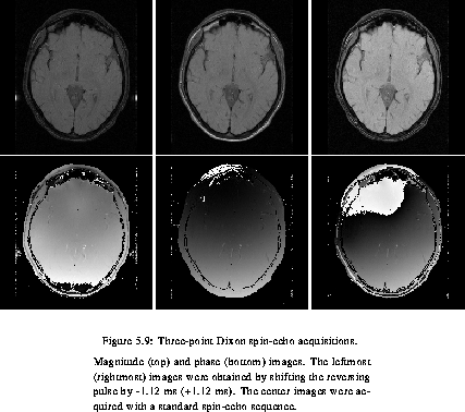
The shifted phase images were unwrapped using the technique described by M. Song [57], who provided us with his computer program.
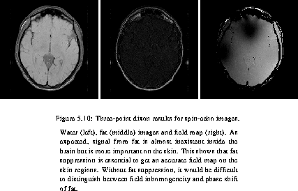
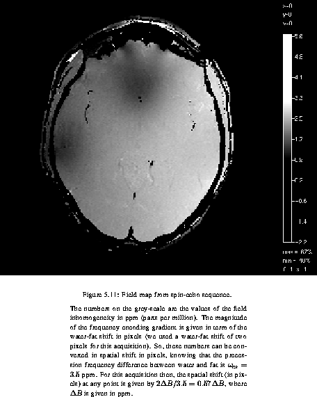
Even though the acquisition of the field map does not require the computation of the water and fat images, their equations have been derived in last section as a means of double checking the field map obtained, since an incorrect field map cannot lead to separation of fat and water. The water, fat and field map images were computed using these equations, and are shown in se_wf. B0map shows the same field map as in se_wf but the gray-scale explicitly relate to the value, in ppm, of the field inhomogeneity displayed.
A similar set of images were acquired using a gradient-echo sequence.
The first was measured with a TE of 11.2 ms and the other two, with a
TE of 8.96 ms and 13.44 ms respectively. Note that the echo time of
the intermediate image is such that  .
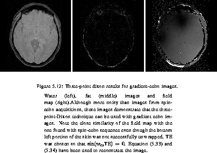
The resulting water, fat and field images are shown in ge_wf.
.
The resulting water, fat and field images are shown in ge_wf.
Observe that the field map actually determined can be affected by eddy currents which are dependant of the MR pulse sequence used. For instance, a gradient-echo sequence with short TE will be affected differently by eddy currents than a spin-echo sequence with long TE.