
MNI Rhesus Macaque Atlas
Overview
We present an unbiased standard rhesus macaque monkey magnetic resonance imaging template brain volume that offers a common stereotaxic reference frame to localize anatomical and functional information in an organized and reliable way for comparison across individual rhesus monkeys and studies.
We have used MRI volumes from a group of 7 normal adult rhesus monkeys (Macaca mulatta) to create the individual atlas. Thus, the atlas does not rely on the anatomy of a single subject, but instead depends on nonlinear normalization of numerous rhesus monkey brains mapped to an average template image that is faithful to the location of anatomical structures.
Tools for registering a native MRI to the rhesus macaque atlas can be found in the Software section.
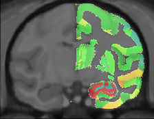
Viewing
Viewing the atlas and associated volumes online requires Java browser support. The Java Internet Viewer (JIV2) used here is available for download and personal use under the GNU general public license (GPL). A custom download of JIV2 and the volumes used below is available here.
- To view the MNI rhesus macaque atlas online: click here.
- To view the MNI rhesus macaque and the Paxinos labels together: click here.
- To view the MNI rhesus macaque along with the Paxinos labels and a sample native rhesus: click here.
The MNI MRI rhesus stereotaxic coordinates (X,Y,Z) are displayed in the first row below the volumes. The second row is reserved for display of the native rhesus coordinates (Xnat,Ynat,Znat), and the third row for displaying the Paxinos atlas coordinates (Breg,Lat,Sup).
For viewing, one can use the left most mouse button to click on any image, and the other cross-sectional images will be updated with the appropriate position. You can also hold the middle/rocker mouse button down while moving up or down, to pan through the image plane. Holding ‘Shift’ with the left or middle button will enable dragging and zooming.
When looking at the images, remember that left is left and right is right!
Methods
The rhesus macaque average atlas is comprised of 7 T1-weighted MRIs of normal young adult rhesus macaque brains. This atlas is not based on a single subject but instead is an average constructed from the averaged position, orientation and scale from all the individual subjects and is representative of both the intensities and spatial positioning of anatomical structures.
The rhesus atlas was created via the macaque atlas (see Macaque for details). By averaging the voxel intensities of the 7 rhesus MRI which were resampled in MNI macaque space, a mean intensity image is created. The deformations for these 7 animals were also averaged together, inverted, and then applied to the mean intensity image to create the unbiased standard rhesus macaque monkey MRI template brain atlas.
Publications
The following publications should be referenced when using this atlas:
- M. Mallar Chakravarty, Stephen Frey, and D. Louis Collins. ‘Digital atlas of the monkey brain in stereotactic co-ordinates’ in The Rhesus Monkey Brain In Stereotactic Coordinates, G Paxinos, XF Huang, M Petrides, AW Toga. Elsevier, March 2008.
- Stephen Frey, Deepak N. Pandya, M. Mallar Chakravarty, Michael Petrides, D. Louis Collins. ‘MNI monkey space’, Neuroscience Research, Volume 65, Supplement 1, pg S130, 2009.
- Stephen Frey, Deepak N. Pandya, M. Mallar Chakravarty, Lara Bailey, Michael Petrides, D. Louis Collins. ‘An MRI based average macaque monkey stereotaxic atlas and space (MNI monkey space)’, NeuroImage (2011), doi:10.1016/j.neuroimage.2011.01.040
Download
Click here to download the atlas and associated files in your chosen format.
Contact
For questions related to the MNI rhesus macaque atlas (rather than the website), contact Stephen Frey