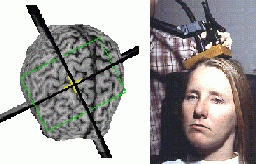
Figure 1: The stimulation of neural tissue in
a subject
|
The 3-Dimensional Mapping of the Magnetic Field |
|
|
| Transcranial Magnetic Stimulators use the principle of electromagnetic induction to produce currents in a conductor by using a moving, or time-varying magnetic field. This phenomena was first described by Micheal Faraday in 1831 at the Royal Institution of Great Britain. In non-invasive magnetic stimulation the stimulating coil acts as one coil space and the electrically conductive living body, as the second coil. This is analogous to the famous Faraday experiment. [1] The ability of magnetic fields to stimulate human tissue was seen as early as 1896 when it was observed that flickering lights in the visual field could be seen when a subject's head was placed in a coil driven by an alternating 110 volt supply at 30 amperes. Facial nerve stimulation was also studied in 1965 by Bickford and Fremming [1], which was followed with the recording of the first muscle evoked potential by Polson et. al. in 1982. The true usefulness of TMS coils was observed in 1985 by Barker et. al. in Sheffield who were able to achieve the first magnetic stimulation of the human motor cortex. [1] |

Figure 1: The stimulation of neural tissue in
a subject
| Since 1985, there has been rapid progression
in this technology. The design of coils is an important area of research
since the varying design of the coil produces different magnetic field
distributions. Coils have been made with multiple turns for accurate
stimulation. The use of train pulses for intraoperative monitoring
and the treatment of depression and other psychiatric disorders are also
part of the scope of a wide range of research being conducted with the
TMS. Magnetic stimulation has also been used to produce fast repetitive
stimuli to determine the laterality of speech centres and high energy stimuli
have been used to restart the fibrillating heart. [1] Figure 1 depicts
a subject who is receiving TMS. The position of the TMS coil on the
subject's brain is shown as two crossing lines which can be determined
with the aid of Magnetic Resonance Images of the subject's brain. (Rogue
Research, Montreal)
In TMS, a brief current is passed through a coil to induce an intense magnetic field. This field in turn, is used to induce a current in human tissue. Depending on the shape and the number of turns in the coil, the magnetic field distribution takes on a unique shape. It is important for a researcher/clinician implementing TMS on his/her patient to know the shape of the magnetic field in order to determine in what areas of the neural tissue currents are being induced. The connectivity and the excitability of the cerebral cortex vary over short distances. It is important to determine the exact position of the coil. Knowing the magnetic field distribution is also useful because it gives the researcher a tool by which he/she can estimate the induced charge density. This is a more useful measure of the effectiveness of the stimulator than just knowing the intensity of the magnetic field strength. The Transcranial Magnetic Stimulating Coil is now being used in the treatment of mood disorders without thorough theoretical understanding of the induced electric/magnetic field distribution. This laboratory exercise is an attempt at bringing us closer to the understanding of the magnetic field and therefore, to a better understanding of the TMS coil and its applications. [2]
|